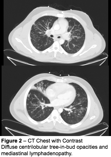
tree in bud opacities causes
The appearance is suggestive of croup which should be obvious clinicallyA corresponding lateral x-ray would show narrowing of the subglottic trachea and ballooning of the hypopharynx. On the left a patient with TB.

Tree In Bud Appearance Radiology Case Radiopaedia Org
Mosaic attenuation is a descriptive term used in describing a patchwork of regions of differing pulmonary attenuation on CT imagingIt is a non-specific finding although is associated with the following.

. The steeple sign also called the wine bottle sign refers to the tapering of the upper trachea on a frontal chest radiograph reminiscent of a church steeple. 2-3 mm nodules with random disrtibution. There is a cavitating lesion and typical tree-in-bud appearance.
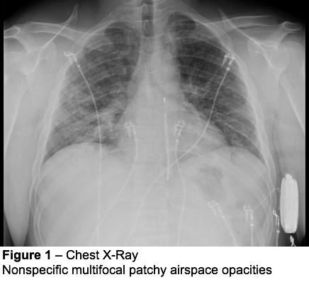
A few COVID-19 cases and findings by dataset. It also results in tree-in-bud opacities and traction bronchiectasis particularly of the upper lobes. A Cardio-vasal shadow within the limits b Increasing left basilar opacity is visible arousing concern about pneumonia c Progressive infiltrate and consolidation d Small consolidation in right upper lobe and ground-glass opacities in both lower lobes e Infection demonstrates right infrahilar airspace opacities and f.
Obstructive small airways disease. They are usually small and associated with parenchymal disease. Air-trapping in lung areas.
The effusions are typically septated and can remain. Low attenuation regions are abnormal and reflect two phenomena occurring at the same time. The blue arrow indicates the biopsy needle.
Pleural Extension Pleural effusions occur most often in primary tuberculosis but are seen in approximately 18 of patients with postprimary tuberculosis. Tree in bud appearance.

Ct Scan Of Chest Revealing Scattered Tree In Bud Opacities In Both Download Scientific Diagram

Tree In Bud Caused By Haemophilus Influenzae Radiology Case Radiopaedia Org

Figure 3 From Tree In Bud Pattern Semantic Scholar

Pdf Tree In Bud Semantic Scholar

Co Rads 2 With Tree In Bud Sign A 27 Year Old Male Attended The Download Scientific Diagram

References In Causes And Imaging Patterns Of Tree In Bud Opacities Chest

It Is Not Always Tuberculosis Tree In Bud Opacities Leading To A Diagnosis Of Sarcoid Abstracts

Hrct Of The Lung Signs Of Infection Bilaterally Tree In Bud Patterns Download Scientific Diagram

Computed Tomography Of The Chest Showed Nodular Opacities With Tree In Download Scientific Diagram

It Is Not Always Tuberculosis Tree In Bud Opacities Leading To A Diagnosis Of Sarcoid Abstracts

Chest Ct With Multifocal Tree In Bud Opacities Diffuse Bronchiectasis Download Scientific Diagram

Signs Of Bronchiectasis Tram Tracks Thick Rings Signet Ring Sign And Finger In Glove Sign This Is Cystic Fibros Medical Ultrasound Radiology Imaging Pet Ct
View Of Tree In Bud The Southwest Respiratory And Critical Care Chronicles

Tree In Bud Caused By Haemophilus Influenzae Radiology Case Radiopaedia Org